1. A cosa serve la provetta per il campionamento dei virus?
Viene utilizzato per la raccolta, trasporto e stoccaggio dell'influenza, influenza aviaria, campioni di coronavirus e altri virus per la successiva estrazione e amplificazione.
2. Qual è la differenza tra soluzioni di conservazione inattivate e non inattivate?
Dopo che i campioni di virus sono stati raccolti, PCR testing cannot be performed in time at the general sample collection site, so the collected virus swab samples need to be transported. The virus will quickly decompose in vitro and affect subsequent testing. Perciò, when storing and transporting, It is necessary to add the virus preservation solution, and for different detection intentions, different virus preservation solutions need to be used. Attualmente, it is mainly divided into two types: inactivated and non-inactivated
Inactivated virus preservation solution:
It is mainly a virus lysis type preservation solution modified by nucleic acid extraction and decomposition solution. Effectively prevent the operator from secondary infection, but it also contains inhibitors, which can prevent the virus nucleic acid from being degraded, and then enable subsequent detection by NT-PCR. And it can be stored at room temperature for a relatively long time, saving the cost of virus sample storage and transportation.
Non-inactivated virus preservation solution:
It is mainly a virus-maintaining liquid-type preservation solution that is improved based on the delivery medium. It can maintain the activity of the virus in vitro and the integrity of the antigen and nucleic acid, maintain the protein coat of the virus from being decomposed, and maintain the originality of the virus sample to a large extent. In addition to nucleic acid extraction and detection, it can also be used for virus cultivation and isolation.
It can be seen that according to different situations, different types of preservation solutions have their own advantages and disadvantages, but it should be noted that whether it is inactivated or non-inactivated, the virus sampling tube before sampling must be strictly inactivated and sterilized to ensure that the tube is inside. There are no other microorganisms, causing the virus to decompose after sampling or other influences causing false detections.
Dopo il prelievo con tampone, if you use poor quality collection tools or preservation solutions, it will affect subsequent test results and even cause false positives for misdiagnosis. Perciò, it is recommended to use good quality virus preservation solutions.
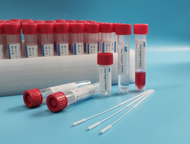
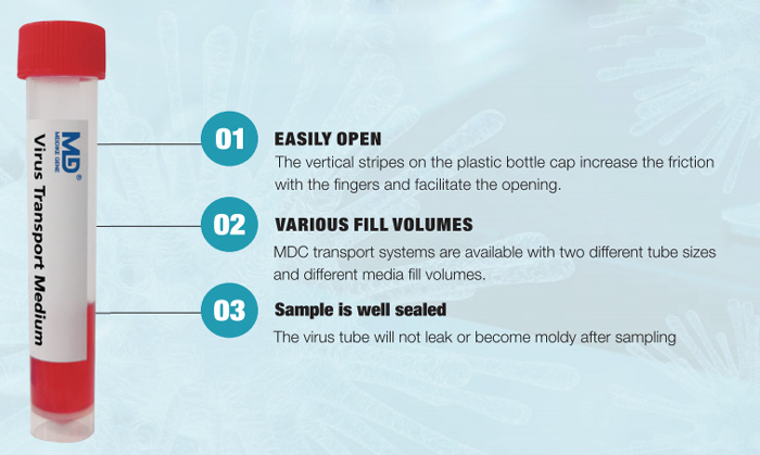
Medico virus sampling tube:

3. How to collect virus Specimen?
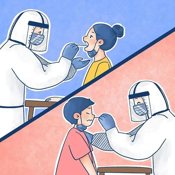
Collection procedures for nasopharyngeal swab and oropharyngeal swab specimen:
Prelievo tampone nasofaringeo
1. The patient’s head is tilted back (circa 70 gradi) and stays still.
2. Use a swab to estimate the distance from the root of the ear to the nostril.
3. Insert from the nostril straight to the face. The deepening interval should be at least half the length of the earlobe to the tip of the nose. After encountering resistance, you should reach the nasopharynx, stay for a few seconds to absorb secretions (generally 15-30s), and rotate the swab 3 A 5 volte.
4. Rotate gently to take out the swab, and immerse the swab head in a collection tube containing 2 mL of lysis solution or a cell preservation solution containing RNase inhibitor.
5. Break off the sterile swab rod at the top, scartare la coda, screw the cap of the tube tightly and close it with a parafilm.

Oropharyngeal swab collection
1. Ask the patient to gargle with saline or water first.
2. Put the swab in sterile normal saline to moisten it.
3. Focus on the patient to sit down, inclinare la testa all'indietro, open his mouth, and make an “ah” suono.
4. Fix the tongue with a tongue depressor. The swab skips the root of the tongue to the posterior pharynx, tonsil recesses, and side walls.
5. First use the swab to scrub the bilateral pharyngeal tonsils back and forth at least 3 volte, and then swallow the posterior wall at least 3 volte, preferably 3 A 5 volte.
6. Take out the swab to prevent touching the tongue, pituitary, oral mucosa and saliva.
7. Immerse the swab head in a preservation solution containing 2 A 3 mL of virus. 8. Break off the sterile swab rod near the top, scartare la coda, screw the tube cap tightly and close it with a parafilm.


